How it works
From a paper ECG to an AI first read
Four steps. The first happens on the phone, the next two inside TraceRead AI, and the last one always belongs to your doctor.
Step 1 of 4
Take the photo
The technician opens the capture app in the phone's web browser, lines the printout up with the guide and takes the photo. Nothing to install.
Photo check
The phone checks the picture about five times a second for blur, glare, poor light and a page that does not fill the frame, and says what to fix.
In testing on 522 frames it flagged every one with blur of half a millimetre or more, large glare, poor light or a page too small, and none of 69 good ones.
Header cover
Before sending, the technician drags a box over the printed header where a machine prints the patient's details.
The box is painted into the photo on the phone, so the covered part never leaves it.
Send when connected
Photos wait safely on the phone and send themselves when the network comes back. Each photo is kept near 1.5 MB.
Each photo travels with a small record of how it was taken, and the server checks its type and size on arrival.
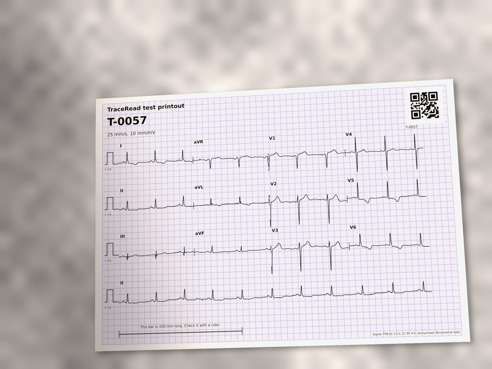
Photo check, tested on 522 frames of TraceRead test printouts with defects added on purpose: 0 of 69 good frames wrongly flagged by the photo check, while it flagged every frame with blur of half a millimetre or more, large glare, poor light or a page too small. TraceRead test S6.
Step 2 of 4
TraceRead AI reads the paper
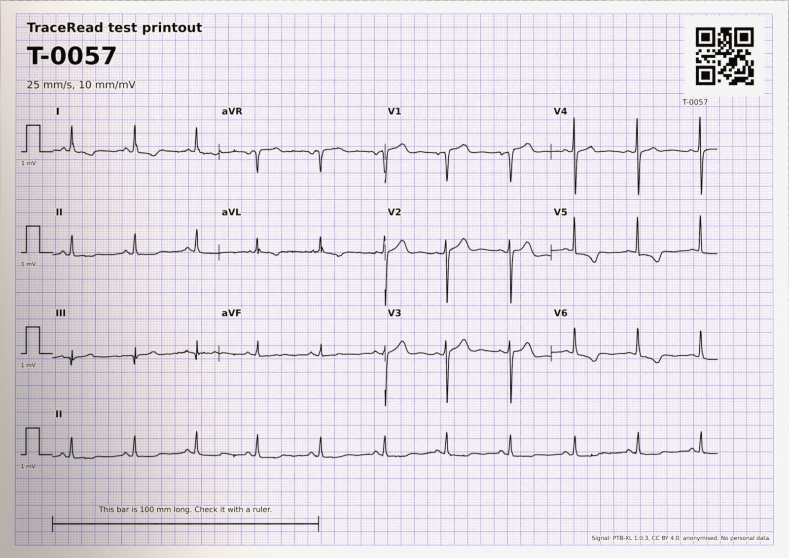
It finds the page, measures the printed grid, reads the layout from the calibration pulses and rebuilds each of the 12 leads as a digital signal.
Page finder
TraceRead AI finds the four corners of the paper and how it is turned, even in a photo taken at an angle.
The printed millimetre grid is then measured to fix the scale and tilt, and to tell A4 from US Letter paper.
Layout reader
TraceRead AI finds the calibration pulse at the start of each row and works out where each of the 12 leads sits on the page.
The pulses are matched against 14 known page layouts. It read the layout right on 99.0% of 500 photos made by computer.
Trace rebuild
TraceRead AI follows the ink of every lead and rebuilds it as a digital signal, 500 points a second.
The rebuilt line is drawn back over the paper. On 500 photos made by computer it matched the original recordings at a median of 0.908.
Coverage map
Marks exactly which seconds of each lead the paper shows, because most printouts show each lead for only 2.5 seconds.
The pattern finder reads the signal and the coverage map together, so a missing part is never treated as a flat line.
Hold for retake
When the rebuilt trace looks unreliable, TraceRead gives no first read and asks for a new photo.
A fixed rule looks at the calibration pulses, the layout match and cut leads. It held 5.9% of 500 photos made by computer.
Step 3 of 4
It measures the tracing and looks for patterns
The measures come from fixed code. The patterns come from TraceRead AI, each with a score from 0 to 100. No generative AI writes anything.
Pattern finder
TraceRead AI checks the tracing for 71 patterns of rhythm, conduction, thickened heart muscle and ST and T changes, each with a score from 0 to 100.
Scored on 2,198 public test ECGs it never trained on (0.902 across all 71), and on 45,152 ECGs from China.
Measures
Heart rate, PR interval, QRS, QT, QTc and heart axis, measured on all the leads together.
Compared with reference ECG programs on 2,198 test ECGs: heart rate within 0.6 beats a minute and QTc within 16.8 ms on average.
- Heart rate
- 63 beats a minute
- PR interval
- 136 ms
- QRS width
- 88 ms
- QT interval
- 406 ms
- QTc, corrected for heart rate
- 414 ms
- Heart axis
- 20 degrees
Patterns and TraceRead AI's score from 0 to 100 (the thin line is where a pattern gets flagged)
Sinus rhythm flagged
95Left ventricular hypertrophy flagged
60Unspecific reduced blood flow pattern flagged
56Reduced blood flow pattern, front and side flagged
27
Step 4 of 4
Your doctor checks and decides
The doctor sees the red rebuilt line over the paper, so a misread lead stands out. They check each number, keep, change or remove each pattern, and decide. Nothing reaches a patient until a doctor has signed it.
An urgent pattern is always written as a question for the doctor, “possible …, review now”, never as a finding.
An AI first read is not the last word.
- A qualified doctor reviews every AI first read. Nothing reaches a patient until a doctor signs it.
- Do not rely on TraceRead alone in an emergency.
- A normal AI first read does not rule out heart disease.
- For adults, 18 and over.
What you need
A phone with a camera and a web browser
The capture app opens from a link. Photos wait on the phone when the network drops.
The printed 12 lead ECG
From an adult, at 25 mm a second and 10 mm per millivolt, laid out as 3x4, 6x2 or 12x1, on A4 or US Letter paper.
A doctor to review
Your own reading doctor checks every AI first read before anything reaches a patient.
See TraceRead read a paper ECG
We show you TraceRead reading public research ECGs, step by step, and talk through how your team prints, photographs and reads ECGs today.